
Your shopping cart is currently empty.
- CRISPR-Cas9
- Cell Culture and Transfection Reagents
- EliteTrust Human Gene ORF Virus Particle Collection
- Featured Products
- Lentiviral Cloning Vectors
- Lentivirus Concentration and Purification kit
- Lentivirus Freeze-Thaw Protection Medium
- Lentivirus Packaging System
- Lentivirus Titration
- Premade Lentivirus
- Premade Stable Cell Line
- Stem Cell Research
- Vectors for iPS Cells Generation
- xpath of 1st category mapping
- xpath of 2nd category mapping
- DNA Mutagenesis
- EliteTrust Custom Adeno-Associated Virus Production
- EliteTrust Custom Gene Targeting Service
- EliteTrust Custom Lentivirus Production
- EliteTrust Custom Retrovirus Production Service
- EliteTrust Custom Stable Cell Line Generation Service
- EliteTrust Fast-Track Cell Authentication Service
- EliteTrust Stable Cell Line Mycoplasma Detection Service
Premade Stable Cell Line
Empowering Discovery with Reporter Stable Cell Line
Cellomics Technology, LLC has developed the world’s largest collections of genetically engineered stable cell lines to accelerate innovation in academic researches and biotechnology and pharmaceutical R&D.
Each of our high-performance cell lines is developed through an evolutionary optimization process — a multi-stage workflow that includes rigorous testing, issue-driven modification, and iterative enhancement — to meet the most demanding research applications. This process takes several months and ensures that the final products deliver:
- Pure, uniform cell populations
- Optimized and stable gene expression levels
- Preserved cellular characteristics
- Precise, application-ready functional responses
The Cellomics premade stable cell line series includes a variety of models tailored for diverse research needs—such as GFP/RFP-labeled cancer cells for in vivo xenograft experiment for animal imaging that can monitor tumor cells growth and metastasis in real-time, CRE-expressing stable cells for gene editing application development, and signaling pathway reporter cell lines for studying the roles of signal transduction pathway in cell behavior.
Cellomics Technology, LLC reporter cell lines includes about more than seven thousands different reporter cell lines, the application of these reporter stable cell lines cover most of common study topics. Stay tuned for more exciting additions as we continue expanding our product portfolio to empower your innovative research.
Among the lots of publications using our stable reporter cell lines, below are two examples:
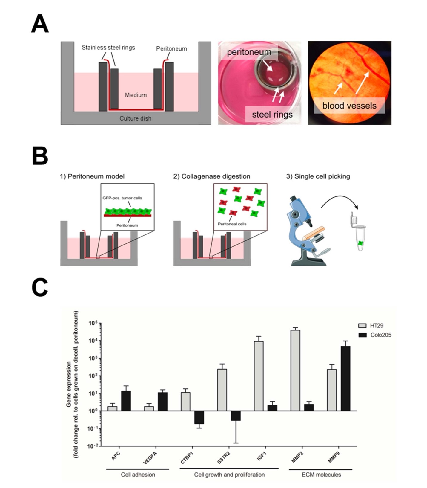
Example 1: Understanding Cancer Invasion in Human Peritoneum
Koch et al., Cancers (2022) 14, 3760. Pharmacologic Targeting of MMP2/9 Decreases Peritoneal Metastasis Formation of Colorectal Cancer in a Human Ex Vivo Peritoneum Culture Model.
In this study, researchers used the Colo205/GFP stable cell line (Cat# SC-1278) engineered by Cellomics Technology, LLC to explore how colorectal cancer cells invade the peritoneum.
The Colo205 cells, expressing strong GFP fluorescence, were seeded on patient-derived peritoneal membranes. After 24 hours, GFP-positive Colo205 cells were isolated directly from the peritoneal layer for gene expression profiling. Using this fluorescence-based selection, scientists successfully captured the early molecular events driving cancer cell invasion, revealed by single-cell next-generation sequencing.
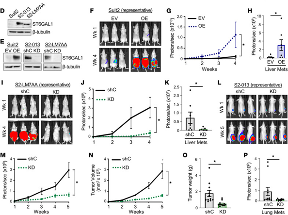
Example 2: Tracking Pancreatic Cancer Progression In Vivo
Bhalerao et al., JCI Insight (2023) 8(19):e161563. ST6GAL1 Sialyltransferase Promotes Acinar-to-Ductal Metaplasia and Pancreatic Cancer Progression.
This study examined the role of ST6GAL1 in pancreatic ductal adenocarcinoma (PDAC) using the Suit2 isogenic series, Suit2, S2-013, and S2-LM7AA, which differ in metastatic potential. Each line was genetically engineered by Cellomics Technology, LLC using a CMV-Firefly Luciferase lentiviral vector (PLV-10003: CMV-Firefly luciferase-PGK-puro) to enable in vivo bioluminescent imaging (BLI). This luciferase expression system allowed scientists to track tumor growth and metastatic spread in real time, providing valuable spatial and temporal data on cancer progression. The BLI approach also revealed metastatic lesions in distant organs — such as liver and lung — that would be difficult to identify by conventional methods. Importantly, tumor growth and metastatic burden measured by BLI were consistent with endpoint tumor size and weight, validating the imaging method’s quantitative accuracy.
Driving Scientific Impact
These examples highlight how Cellomics Technology’s engineered stable cell lines empower cutting-edge research on dissecting molecular mechanisms of cancer invasion by enabling real-time tracking of tumor dynamics in vivo.
Stable cell lines are invaluable tools for biomedical research and drug discovery, offering consistent and reproducible models for studying complex biological processes. Cellomics’ premade stable cell lines are generated using our well-established lentiviral platform and selected through antibiotic resistance. Because lentiviruses integrate stably into the host genome after transduction, these cell lines continuously express the target genes across subcultures.
All the premade stable cell lines can be searched by either the names of cell line, reporter, or pathway. In case the cell line name has just two characters such as "EA", please then search for "EA cell". Below please find the available most common cell lines and reporters and pathways covered in our collection:
| Cell Line Names
|
Reporter/Pathways/Imaging
|
The above products can also be browsed through the below five categories.